
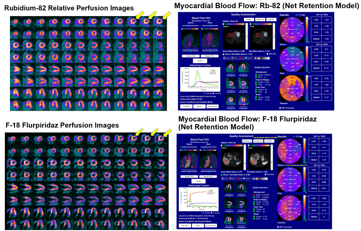
 “Serial scans are now increasingly performed in cardiac transplant patients as a part of CAV surveillance. This case illustrates how radiotracer selection can impact what you see.”
– James A. Case, PhD, MASNC
Author of Visual Perfusion vs Absolute Blood Flow: Differences in Imaging Agents
How much of what you see on a PET myocardial perfusion image reflects the underlying disease, and how much reflects the properties of the radiotracer?
New case on ASNC.org! Visual Perfusion vs Absolute Blood Flow: Differences in Imaging Agents prompted this month’s Cardiac PET Case Challenge. We asked members to review the patient’s perfusion images and myocardial blood flow findings and vote on what their interpretation suggests about CAV progression. View the poll.
This month’s cardiac PET case explores this question through serial PET studies in a cardiac transplant patient undergoing surveillance for coronary allograft vasculopathy. The case compares studies performed one year apart using different PET radiotracers and illustrates why differences in image appearance do not necessarily indicate disease progression.
Article Type
News & Announcements
Category
Education
Related Posts

JNC Editor Challenges Nuclear Cardiology Community to Advance Inflammation Imaging and Clinical Evidence
Advancing molecular imaging of myocardial inflammation will require a concerted effort from…
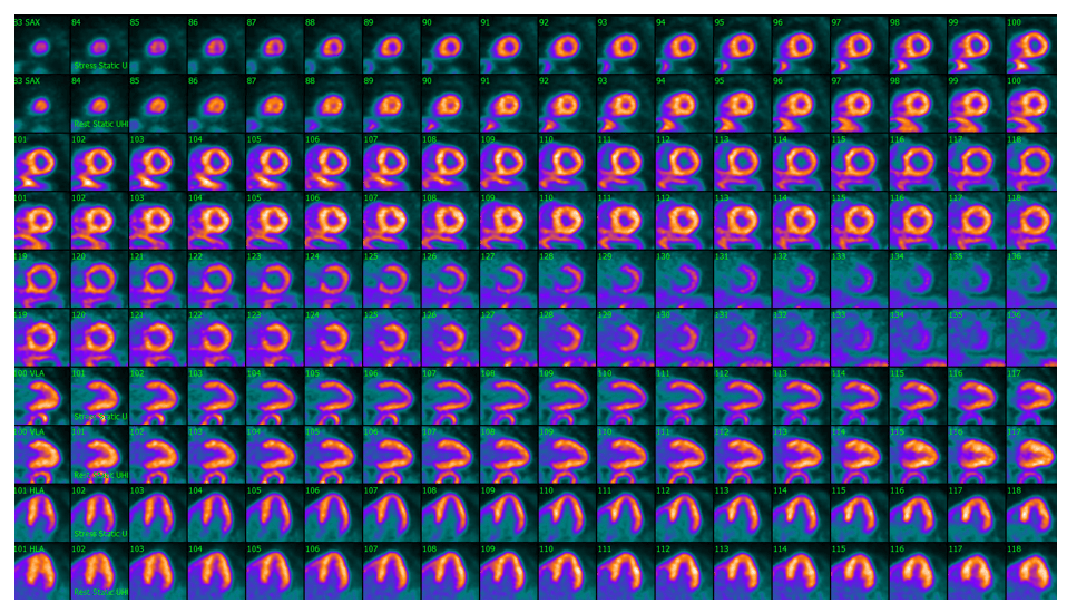
Cardiac Amyloidosis and Suspected Coronary Disease: What Would You Do Next?
An 80-year-old man with chest pain and known ATTR cardiac amyloidosis, diagnosed…

CMS Releases Final Medicare Physician Fee Schedule and Hospital Outpatient Payment Rates and Policies for 2027
The Centers for Medicare & Medicaid Services (CMS) has released the 2027…