
ASNC offers a variety of courses, meetings, programs, and products covering the latest developments in nuclear cardiology and multimodality imaging to meet the needs of our members and other professionals in the field. Our educational programming is at the core of ASNC’s mission to foster quality nuclear cardiology services that provide the utmost in patient care.
Each of ASNC’s educational activities is planned and developed with oversight and input from our Education Committee. Many of our activities offer Continuing Medical Education (CME) credits, medical knowledge Maintenance of Certification (MOC) points, and Continuing Education (CE) credits.
ASNC is proud to have received the highest level of recognition Accreditation with Commendation by the Accreditation Council for Continuing Medical Education (ACCME) through July 31, 2029.
ASNC Events
Learn from experts in the field at one of ASNC’s upcoming events offered in person, virtually live, or OnDemand.
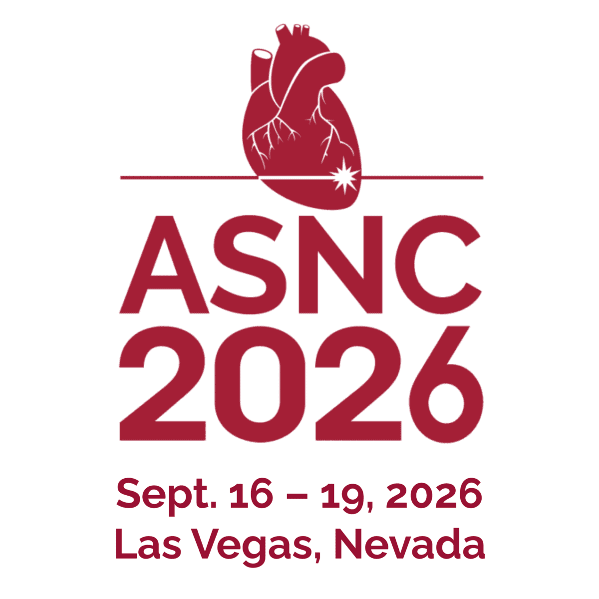
ASNC2026 Annual Scientific Session & Exhibition
Sept. 16-19, 2026 • Las Vegas
Register Now and Save Up to $200 with Early Registration Ratess!

You are invited to join us for the 31st Annual Scientific Session and Exhibition of the American Society of Nuclear Cardiology, taking place Sept. 16-19, 2026, at Caesars Palace in iconic Las Vegas, Nevada.
In-person and virtual registration are available.
Bonus! The Imaging Techniques and Standards virtual program on Sept. 26 is included free with your registration, offering additional focused education you can access from anywhere.

ASNC offers a robust portfolio of CME/CE/MOC to help you meet your goals to increase your knowledge, skills, and professional performance in the field.
Sponsorship & Advertising
Your organization can be a part of the continuing evolution in nuclear cardiology by collaborating with ASNC to support your customers’ success. Take advantage of our comprehensive portfolio of opportunities to help you achieve your goals and enhance relationships with imaging professionals.

Earn nuclear cardiology
AMA PRA Category 1 Credits™,
ARRT Category A Credits, and medical knowledge Maintenance of Certification (MOC) points in a variety of topics including cardiac amyloidosis, cardiac PET, and hybrid imaging.
Document your facility’s commitment to quality and safety through lab accreditation.
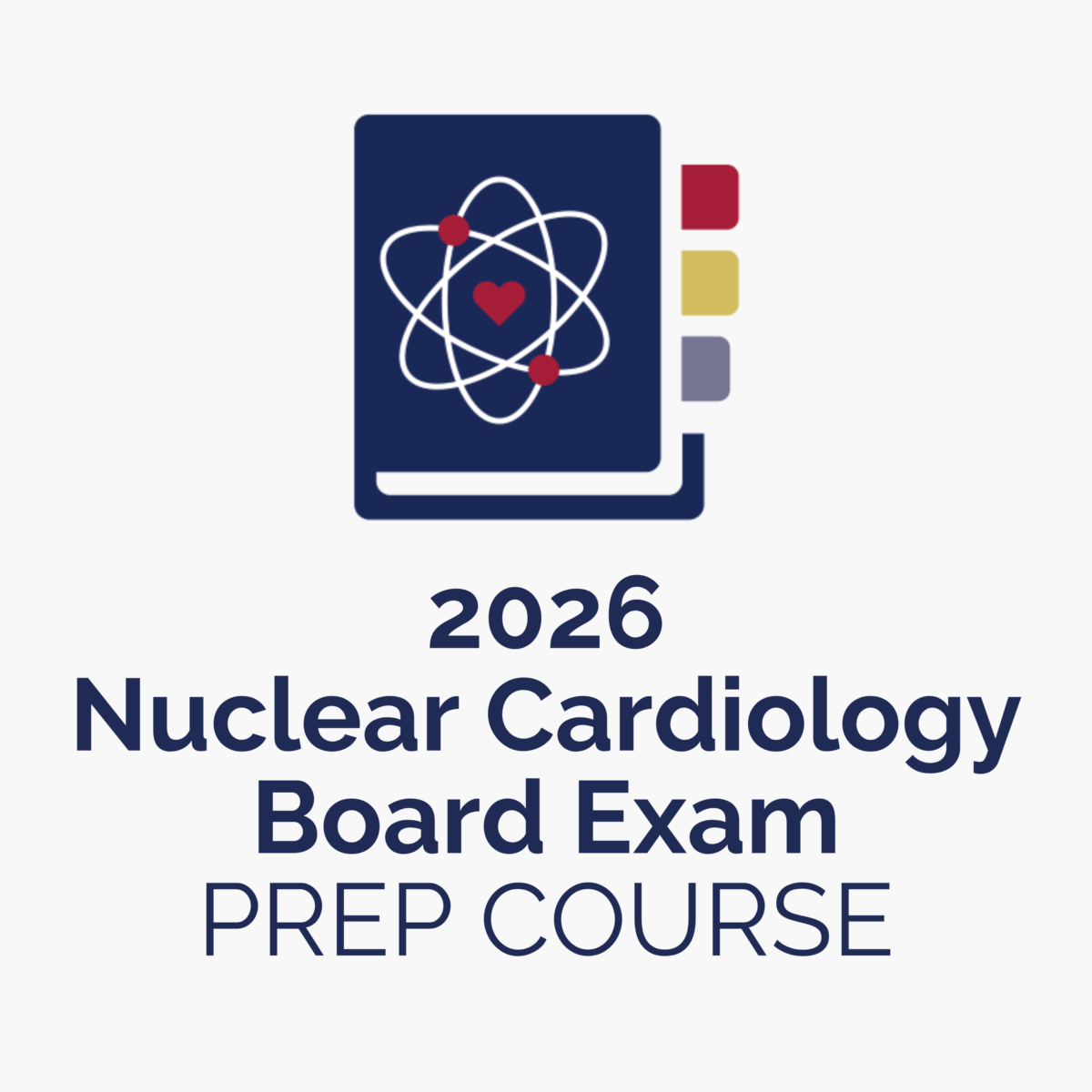
Plan for the CBNC Board Exams with the leading nuclear cardiology board prep course.
